Microfossil Modeling with MicroCT
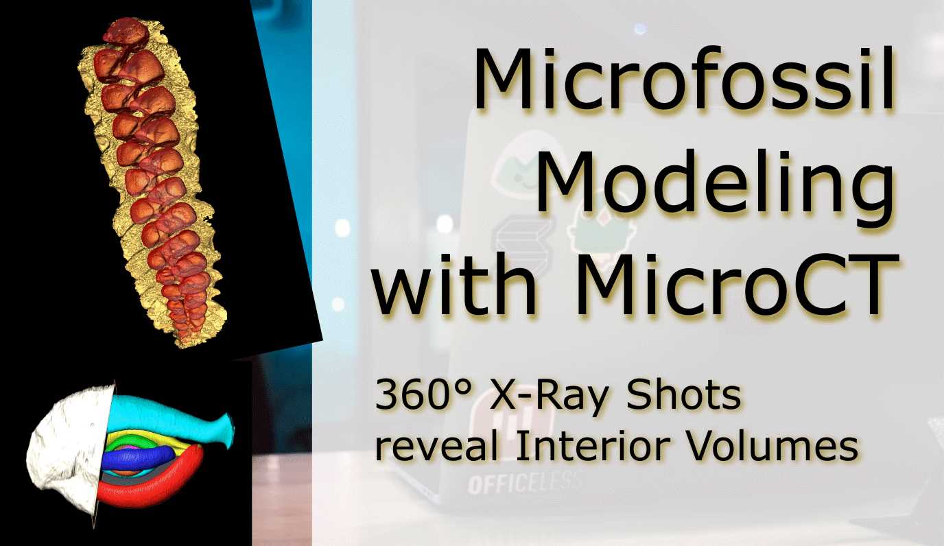
360° X-Ray Shots reveal Interior Volumes
Introduction
The MicroCT housed at the Steinmann Institut für Geowissenschaften can scan specimens from large to small (think Brachiosaur skull and friends).
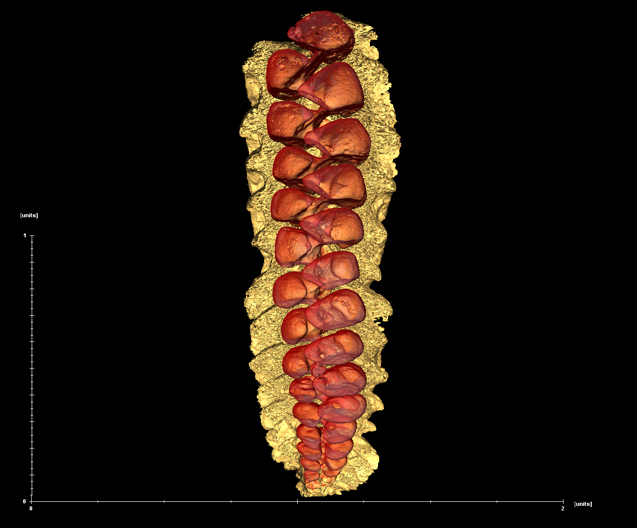
With a lower resolution limit of 2 micrometers, it is possible to get satisfactory details on microfossils, such as foraminifera in the size range 1-2 millimeters.
The Langer Lab deviced a new mounting technique to successfuly scan foraminifera and reconstruct digital 3D models from tomography images.
Goal is measuring structures and volumes of foraminiferal shells without destroying precious specimens.
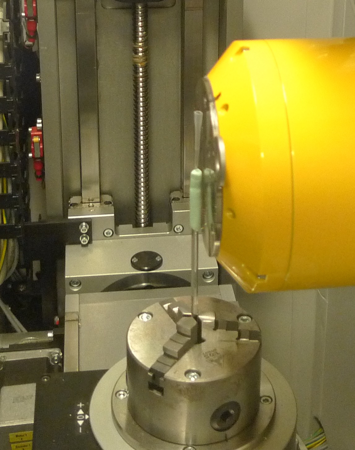
In the figure above the yellow cylinder on the right hand side is the X-Ray source.
The specimen is mounted in the hollow plastic cone (pipette tip) directly in front of the opening of the source.
The single foraminifera (~1 millimeter) is too small to be seen from this distance.
Recorded are the images on a CCD sensor on the left (not pictured) similar to the device in a digital camera, but much larger.
The specimen is rotaded 360 degrees in front of the source and images taken at each degree.
After the recording is finished, the computer will calculate a black-and-white image stack that is imported into a modeling suite (“reconstruction”).
The 3D model can be manipulated and specific materials and volumes marked and measured (see linked materials below).